The Cerebrum
|
|
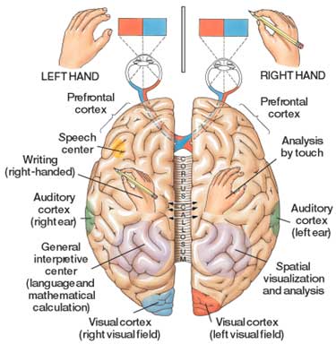
The cerebrum is the largest part of the brain where conscious thought, awareness, feelings, ideas, decision making, and memories occur. Voluntary movements, movements that you are aware of and can start and stop, are generated and controlled in this part of the brain. It accounts for four fifths of the brain’s total weight and 85%-90% of the forebrain. The cerebrum is divided into two cerebral hemispheres, a left hesmisphere and a right hemisphere. Between the hemispheres is the corpus callosum, a bridge, that connects them. The corpus callosum consists of more than 200 million nerve fibers that carry nerve signals between the two halves of the brain so that each half knows what the other half is doing. Each hemisphere is divided into three main parts: the cerebral cortex, cerebral medulla, and basal ganglia (Farley et al., 2014) (Roca & Serrano, 1996) (Olesky, 2001) (Parker, 1997) (Walker & Wood, 2003).
The cerebral cortex is the wrinkled, shiny, grayish pink outer layer of the cerebrum. Cortex is the Latin word for “bark”. It is approximately 0.16 inches think and if unfolded and spread out, it would cover the top of an office desk. Here 50 billion nerve cells link together with trillions of dendrites form a huge network of pathways for nerve signals. The cerebral cortex is divided into different centers where different senses and functions are concentrated. Centers are generally in pairs with one on each cerebral hemisphere. Each center works with the opposite side of the body. For example the visual center in the left hemisphere works with the right eye. Large parts of the cortex have no known functions. Scientists think that these areas deal with awareness, thoughts, feelings, decisions, and memory (Farley et al., 2014) (Roca & Serrano, 1996) (Olesky, 2001) (Parker, 1997) (Walker & Wood, 2003).
The inner cerebral medulla is located below the cortex. It is the white matter of the brain due to its color as a result of all the myelin surrounding the axons contained there. The axons connect the basal ganglia to the other parts of the brain. The basal ganglia are the lobes and curved lumps detected by the wrinkled appearance of the folded surface of the brain. The bulges are called gyri and the grooves are called sulci. Extra deep groves called fissures divide each hemisphere into lobes or main patches. The four lobes are named for the curved bones of the skull around them. The lobes of the brain are the:
Frontal Lobe - involves reasoning planning speech movement emotions problem solving
Parietal Lobe handles sensory input including pain, heat, cold, touch, taste, and pressure.
Temporal Lobe - processes sound and relays information to the parietal and frontal lobe.
Occipital Lobe - receives and processes visual information and sends it to the frontal and parietal lobes.
(Farley et al., 2014) (Roca & Serrano, 1996) (Olesky, 2001) (Parker, 1997) (Walker & Wood, 2003).
